Upper Back Muscles Diagram : Upper Back Muscle Anatomy Anatomy Drawing Diagram - Muscles of the back diagram.. Lower back muscle diagram anatomy does degenerative disc disease affect the lower back muscle? Rows target the muscles of your upper back and back of your shoulder. These structures work together to support the body, enable a range of movements, and send messages from the brain to. It is very stiff, and the thoracic spine has a limited range of motion. Other muscles that aid in shoulder movement include:
Together, these muscles straighten your knee, stabilize your knee joint, assist in flexing your hip (drawing your knee towards your chest), and help absorb force when you land after jumping or leaping. This is a diagram of the larger and more surface muscles of the low back. Now take your left hand and interlace it around the right arm. Keep your elbows bent at a 90 degree angle. There are three sets of longissimus muscles:
Build Upper Back Muscles Off 74 from image.boxrox.com The upper back is the area between the base of the neck and the bottom of the ribcage. Now take your left hand and interlace it around the right arm. Rows target the muscles of your upper back and back of your shoulder. There are 12 bones that make up the upper back, which doctors call the thoracic spine. The longissimus (red, in the image above) are located between spinalis and the iliocostalis muscles. Here are 10 of the best upper back exercises to get you started. Flexes elbow and moves forearm. These structures work together to support the body, enable a range of movements, and send messages from the brain to.
1) above the cervical area (longissimus capitis), 2) in the cervical area (longissimus cervicis), and 3) in the upper back or thoracic area (longissimus thoracis).
Together, these muscles straighten your knee, stabilize your knee joint, assist in flexing your hip (drawing your knee towards your chest), and help absorb force when you land after jumping or leaping. Neck muscles are bodies of tissue that produce motion in the neck when stimulated. You use this muscle when you stand up, walk, run, and climb stairs in fact—whenever you straighten or extend your legs. Listed below are common areas of pain, or you can download a copy here. The four muscle groups that together make up the deep muscle group are the segmental muscles, the transversospinales, the erector spinae, and the spinotransversales. Upper back, lower back, lats, traps, spinal erectors—the whole deal. The longissimus (red, in the image above) are located between spinalis and the iliocostalis muscles. The muscles of the chest and upper back occupy the thoracic region of the body inferior to the neck and superior to the abdominal region and include the muscles of the shoulders. The back consists of the spine, spinal cord, muscles, ligaments, and nerves. The quad muscles— which form the meaty mass on the front of your thighs — are among your strongest muscle groups, and play a critical role in athletic activities. 1) above the cervical area (longissimus capitis), 2) in the cervical area (longissimus cervicis), and 3) in the upper back or thoracic area (longissimus thoracis). 7 stretches for upper back, neck and shoulders: Learn vocabulary, terms, and more with flashcards, games, and other study tools.
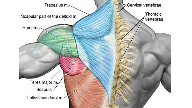
Both the deltoid and the trapezius are firmly attached to the spine of the scapula. We think this is the most useful anatomy picture that you need. This is a diagram of the larger and more surface muscles of the low back. The quad muscles— which form the meaty mass on the front of your thighs — are among your strongest muscle groups, and play a critical role in athletic activities. Together, these muscles straighten your knee, stabilize your knee joint, assist in flexing your hip (drawing your knee towards your chest), and help absorb force when you land after jumping or leaping.
Training Upper Back Off 62 from i.pinimg.com For more anatomy content please follow us and visit our website: The upper arm is located between the shoulder joint and elbow joint. It is like that for several reasons, all of which you can understand by looking at the anatomy of the thoracic spine. To learn more about the anatomy of the spine, watch this video. The muscles of the back are a group of strong, paired muscles that lie on the posterior aspect of the trunk they provide movements of the spine, stability to the trunk, as well as the coordination between the movements of the limbs and the back muscles are divided into two large groups: And the science backs it up. Adductors (includes madductor longus, adductor brevis, adductor magnus muscles: Together with several other muscles, the gluteus maximus muscles form the buttocks.
The upper arm is located between the shoulder joint and elbow joint.
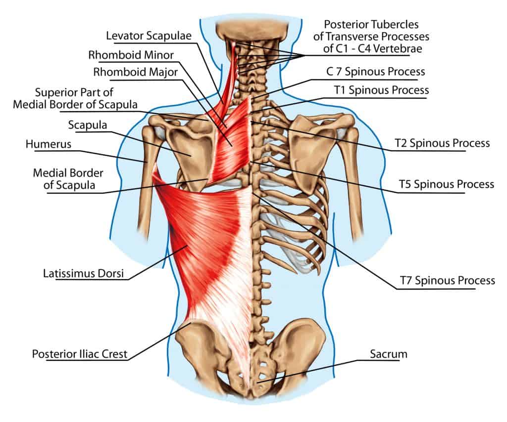
We hope this picture anatomy of back muscles diagram can help you study and research. The deltoid, teres major, teres minor, infraspinatus, supraspinatus (not shown) and subscapularis muscles (not shown) all extend from the scapula to the humerus and act on the shoulder joint. For optimum maximum muscle contraction, squeeze the shoulder blades together at the end of each pull, before releasing back to the front. These important muscles control many motions that involve moving the arms and head — such as throwing a ball, looking up at the sky, and raising your hand. Together, these muscles straighten your knee, stabilize your knee joint, assist in flexing your hip (drawing your knee towards your chest), and help absorb force when you land after jumping or leaping. These structures work together to support the body, enable a range of movements, and send messages from the brain to. 1) above the cervical area (longissimus capitis), 2) in the cervical area (longissimus cervicis), and 3) in the upper back or thoracic area (longissimus thoracis). The spinal erecotrs allow you to flex and extend your back in any given direction. The trapezius, rhomboid and levator muscles of the shoulder. There are three sets of longissimus muscles: Most of the time, back muscle pain is diagnosed then treated with little more than a prescription of rest, painkillers and muscle relaxants. The upper arm is located between the shoulder joint and elbow joint. Neck muscles are bodies of tissue that produce motion in the neck when stimulated.
Together with several other muscles, the gluteus maximus muscles form the buttocks. For optimum maximum muscle contraction, squeeze the shoulder blades together at the end of each pull, before releasing back to the front. The upper back is the area between the base of the neck and the bottom of the ribcage. The upper arm is located between the shoulder joint and elbow joint. The muscles of the back are a group of strong, paired muscles that lie on the posterior aspect of the trunk they provide movements of the spine, stability to the trunk, as well as the coordination between the movements of the limbs and the back muscles are divided into two large groups:
The Complete Guide To Upper Body Muscles For Beginners Empower Your Wellness from empoweryourwellness.online The gluteus maximus is the largest muscle in the body. The extrinsic (superficial) back muscles, which lie most superficially on the back. It is like that for several reasons, all of which you can understand by looking at the anatomy of the thoracic spine. There are 12 vertebrae in the thoracic spine. The spinal erecotrs allow you to flex and extend your back in any given direction. Learn vocabulary, terms, and more with flashcards, games, and other study tools. Rows target the muscles of your upper back and back of your shoulder. This is my video about the muscles of the back.
This is a great stretch to release tight trigger points in between your shoulder blades.
Lower back muscle diagram anatomy does degenerative disc disease affect the lower back muscle? This is a great stretch to release tight trigger points in between your shoulder blades. This move works your upper back muscles, as well as your shoulders and core, friedman says. The upper back is the area between the base of the neck and the bottom of the ribcage. Holding a dumbbell in each hand, start in a high plank position with your wrists under your shoulders and your head, hips, and heels in a straight line. This is a diagram of the larger and more surface muscles of the low back. Learn vocabulary, terms, and more with flashcards, games, and other study tools. There are 12 bones that make up the upper back, which doctors call the thoracic spine. Musculoskeletal, shoulder & back back muscles. Select a muscle group under each area to see the corresponding trigger points, referred pain patterns and stretches that should be performed along with pressure pointer treatment. #back muscles chart #back muscles diagram and ligaments #back muscles diagram lats #back muscles diagram massage #upper back muscles chart related posts of back muscles chart muscles of lower leg with base of pelvis The longissimus (red, in the image above) are located between spinalis and the iliocostalis muscles. The trapezius, rhomboid and levator muscles of the shoulder.